The anatomy of flowering plants, or angiosperms, reveals a fascinating organization of cells and tissues that work in harmony to sustain life and reproduction. Among the critical components of a plant, leaves stand out as vital organs primarily responsible for photosynthesis, the process that converts light energy into chemical energy to fuel plant growth. In angiosperms, leaves exhibit remarkable structural diversity, particularly between monocotyledons (monocots) and dicotyledons (dicots). One of the most distinguishing features is the internal structure of their leaves, with dorsiventral leaves being characteristic of dicots and isobilateral leaves typical of monocots.
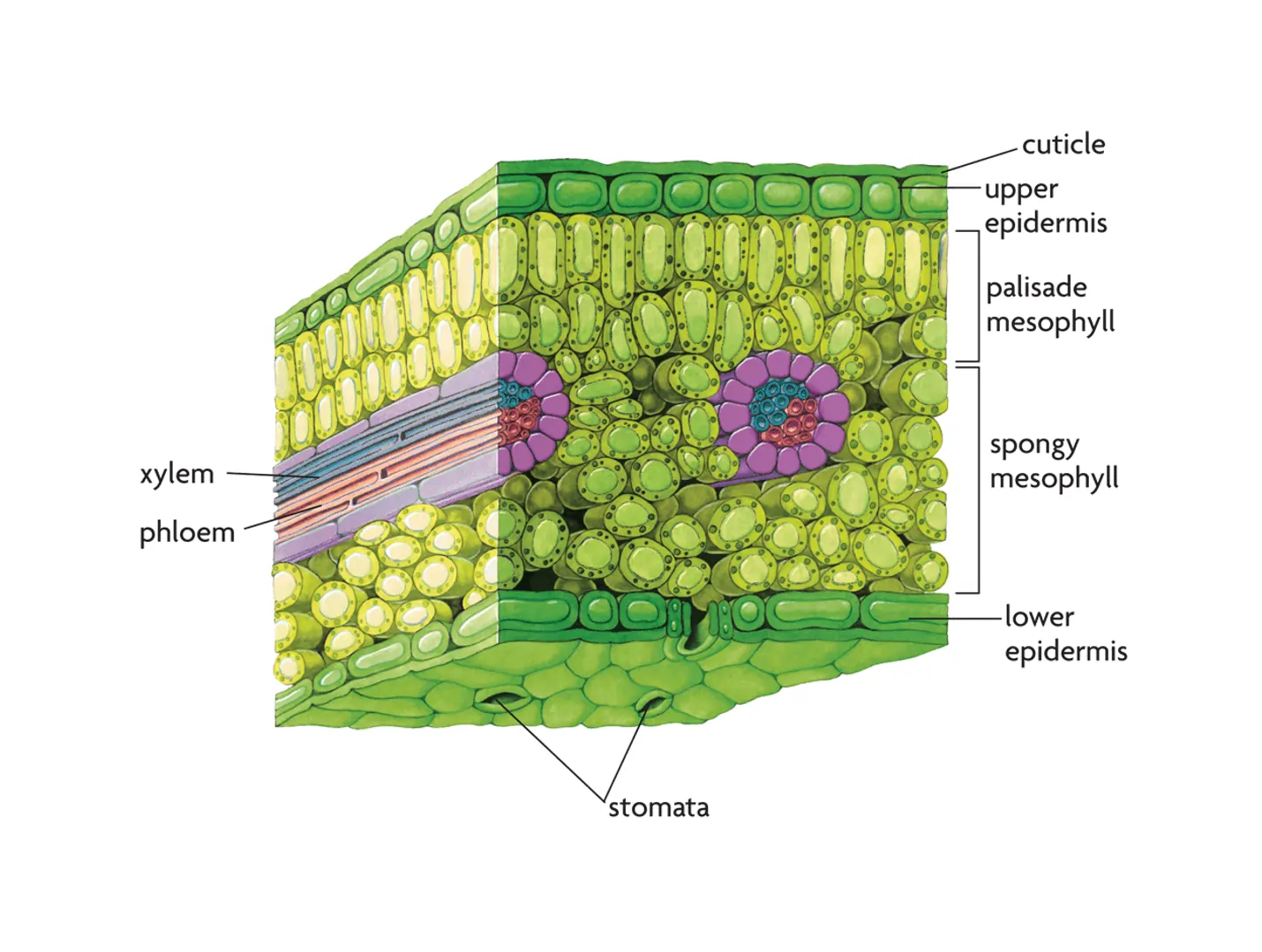
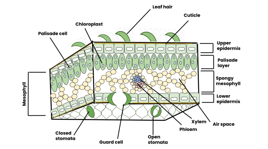
This article delves deeply into the internal structure of a dorsiventral leaf, exploring its components, their functions, and their significance in plant physiology. By examining the upper epidermis, lower epidermis, mesophyll, and vascular bundles, we uncover the intricate design that enables leaves to perform their life-sustaining roles.
Table of Contents
Overview of Angiosperm Leaves and Their Importance
Leaves are often described as the “kitchen” of plants, where photosynthesis occurs, producing glucose and oxygen from carbon dioxide and water using sunlight. Beyond photosynthesis, leaves facilitate gas exchange, transpiration, and sometimes even storage or defense. The internal structure of a leaf is a marvel of evolutionary adaptation, tailored to maximize efficiency in these functions. In dicotyledonous plants, leaves are typically dorsiventral, meaning they have distinct upper (adaxial) and lower (abaxial) surfaces, each with specialized tissues that contribute to the leaf’s overall functionality. In contrast, monocotyledonous leaves are often isobilateral, with similar structures on both sides, reflecting their adaptation to different environmental conditions.

The dorsiventral leaf is a hallmark of dicots, such as beans, roses, or oak trees. Its internal structure is organized into three primary tissue systems: the dermal tissue (epidermis), the ground tissue (mesophyll), and the vascular tissue (vascular bundles). Each of these systems plays a specific role, and their arrangement is optimized for efficient photosynthesis, gas exchange, and nutrient transport. To fully appreciate the complexity of a dorsiventral leaf, we must explore each component in detail, supported by examples and additional insights into their biological significance.
The Dermal Tissue System: Upper and Lower Epidermis
The dermal tissue system forms the outermost protective layers of the leaf, acting as a barrier against environmental stresses such as desiccation, pathogens, and physical damage. In a dorsiventral leaf, this system consists of the upper epidermis and the lower epidermis, each with distinct characteristics that reflect their roles in the leaf’s function.
Upper Epidermis: The Protective Shield
The upper epidermis is a single layer of tightly packed parenchymatous cells located on the adaxial surface of the leaf, which is typically exposed to sunlight. These cells are generally rectangular or tabular in shape and lack intercellular spaces, ensuring a compact barrier. The outer walls of these cells are coated with a waxy layer called the cuticle, which minimizes water loss through transpiration and protects against UV radiation and pathogens. The thickness of the cuticle varies depending on environmental conditions—thicker in arid environments and thinner in humid ones.
A notable feature of the upper epidermis is the presence of fewer stomata compared to the lower epidermis. Stomata are microscopic pores flanked by specialized guard cells that regulate gas exchange and water vapor loss. In many dicot species, such as sunflowers (Helianthus annuus), the upper epidermis may have only a small number of stomata or none at all, as the primary function of this layer is protection rather than gas exchange. The absence of chloroplasts in most upper epidermal cells further emphasizes their non-photosynthetic role, although some transparent cells may allow light to penetrate to the underlying photosynthetic tissues.
Lower Epidermis: The Gateway for Gas Exchange
In contrast, the lower epidermis, located on the abaxial surface, is designed to facilitate gas exchange. Like the upper epidermis, it consists of a single layer of parenchymatous cells covered by a cuticle, but it typically contains a significantly higher number of stomata. These stomata are critical for allowing carbon dioxide to enter the leaf for photosynthesis and oxygen to exit, as well as regulating water vapor loss during transpiration. The guard cells surrounding each stoma contain chloroplasts, enabling them to respond to light and control the opening and closing of the stomatal pore.
For example, in plants like tobacco (Nicotiana tabacum), the lower epidermis may have 100–300 stomata per square millimeter, compared to 0–50 on the upper epidermis. This distribution reflects the adaptation of dorsiventral leaves to maximize gas exchange while minimizing water loss on the sun-exposed upper surface. The lower epidermis’s role in gas exchange is further supported by the spongy parenchyma beneath it, which contains air spaces that enhance the diffusion of gases to and from the stomata.
Table: Comparison of Upper and Lower Epidermis in a Dorsiventral Leaf
| Feature | Upper Epidermis | Lower Epidermis |
|---|---|---|
| Location | Adaxial (upper) surface | Abaxial (lower) surface |
| Stomata Density | Low (few or none) | High (numerous stomata) |
| Cuticle Thickness | Thicker | Thinner |
| Chloroplasts | Absent in most cells | Present in guard cells |
| Function | Protection, light penetration | Gas exchange, transpiration |
| Cell Arrangement | Tightly packed, no intercellular spaces | Tightly packed, no intercellular spaces |
This table illustrates the structural and functional differences between the two epidermal layers, highlighting their complementary roles in the dorsiventral leaf.
The Ground Tissue System: Mesophyll
The mesophyll is the ground tissue located between the upper and lower epidermis, forming the bulk of the leaf’s internal structure. In dorsiventral leaves, the mesophyll is differentiated into two distinct regions: the palisade parenchyma and the spongy parenchyma. This differentiation is a key characteristic that distinguishes dorsiventral leaves from the isobilateral leaves of monocots, where such specialization is typically absent.
Palisade Parenchyma: The Photosynthetic Powerhouse
The palisade parenchyma is located just beneath the upper epidermis and consists of elongated, cylindrical parenchymatous cells arranged in a tightly packed, columnar fashion. These cells are rich in chloroplasts, the organelles responsible for photosynthesis, making the palisade parenchyma the primary site of light capture and energy production. The elongated shape and vertical orientation of these cells maximize their exposure to sunlight penetrating through the upper epidermis, enhancing photosynthetic efficiency.
In plants like pea (Pisum sativum), the palisade parenchyma may consist of one to three layers of cells, depending on the species and environmental conditions. The cells have minimal intercellular spaces, which ensures a high density of chloroplasts and efficient use of space for photosynthesis. The chloroplasts contain chlorophyll, the pigment that absorbs light energy, particularly in the blue and red wavelengths, driving the synthesis of glucose.
Spongy Parenchyma: Facilitating Gas Exchange and Flexibility
Beneath the palisade parenchyma lies the spongy parenchyma, a layer of loosely arranged, irregularly shaped cells with large intercellular spaces. These air spaces are a defining feature of spongy parenchyma, creating a network of channels that facilitate the diffusion of gases (carbon dioxide, oxygen, and water vapor) between the mesophyll and the stomata in the lower epidermis. The cells of the spongy parenchyma also contain chloroplasts, but in lower numbers than the palisade parenchyma, indicating a secondary role in photosynthesis.
The spongy parenchyma’s structure is well-suited for gas exchange, as the air spaces allow rapid diffusion of carbon dioxide to the palisade parenchyma for photosynthesis and oxygen out of the leaf. In plants like Mangifera indica (mango), the spongy parenchyma may also serve as a storage site for water or nutrients, providing flexibility to the leaf under varying environmental conditions. The irregular arrangement of cells also contributes to the leaf’s structural integrity, allowing it to withstand mechanical stresses such as wind or rain.
Table: Characteristics of Palisade and Spongy Parenchyma
| Feature | Palisade Parenchyma | Spongy Parenchyma |
|---|---|---|
| Location | Beneath upper epidermis | Beneath palisade parenchyma, near lower epidermis |
| Cell Shape | Elongated, cylindrical | Irregular, round or oval |
| Intercellular Spaces | Minimal | Large, abundant |
| Chloroplast Density | High | Moderate to low |
| Primary Function | Photosynthesis | Gas exchange, some photosynthesis |
| Arrangement | Tightly packed, columnar | Loosely arranged |
This table highlights the complementary roles of the two mesophyll layers, with the palisade parenchyma optimized for photosynthesis and the spongy parenchyma facilitating gas exchange.
The Vascular Tissue System: Vascular Bundles
The vascular tissue system forms the “circulatory system” of the leaf, responsible for transporting water, nutrients, and photosynthetic products. In a dorsiventral leaf, this system is organized into vascular bundles, which are embedded within the mesophyll and visible as the veins of the leaf. The vascular bundles consist of two main types of tissues: xylem and phloem, each with specialized functions.
Structure and Arrangement of Vascular Bundles
The vascular bundles are arranged in a reticulate venation pattern, a characteristic feature of dicot leaves, where veins form a branching network. The largest vascular bundle, often referred to as the midrib, runs centrally through the leaf, while smaller veinlets branch out to supply the mesophyll. Each vascular bundle is surrounded by a layer of bundle sheath cells, which are typically parenchymatous and provide structural support and sometimes additional photosynthetic capacity.
Within the vascular bundle, the xylem is oriented toward the upper epidermis and is responsible for transporting water and minerals from the roots to the leaf. The phloem, located toward the lower epidermis, transports the products of photosynthesis, such as sugars, to other parts of the plant. The xylem consists of tracheids and vessel elements for water conduction, while the phloem contains sieve tubes and companion cells for nutrient transport. In species like Rosa indica (rose), the vascular bundles vary in size, with the midrib being the largest and the veinlets progressively smaller, ensuring efficient distribution of resources throughout the leaf.
Functional Significance of Vascular Bundles
The vascular bundles form the structural skeleton of the leaf, providing mechanical support to maintain its shape. They also ensure that the photosynthetic tissues receive a steady supply of water and minerals, which are essential for photosynthesis and other metabolic processes. The reticulate venation pattern allows for redundancy in the transport system, ensuring that if one vein is damaged, others can still supply the leaf. For example, in oak leaves (Quercus robur), the robust vascular network supports the leaf’s large surface area, enabling it to withstand environmental stresses while maintaining efficient resource transport.
Additional Insights: Adaptations and Variations
The internal structure of a dorsiventral leaf is not static; it varies across species and environments, reflecting adaptations to specific ecological niches. For instance, in xerophytic dicots like Nerium oleander (oleander), the upper epidermis may have a thicker cuticle and sunken stomata to reduce water loss in arid conditions. Similarly, the mesophyll of shade-tolerant plants, such as ferns or certain understory dicots, may have a higher proportion of spongy parenchyma to maximize gas exchange in low-light environments.
Another fascinating adaptation is the presence of specialized cells in the mesophyll, such as crystal-containing idioblasts or mucilage cells, which store water or deter herbivores. In some dicots, like Begonia, the mesophyll may also contain collenchyma or sclerenchyma cells near the veins, providing additional mechanical support. These variations highlight the versatility of the dorsiventral leaf structure in meeting the diverse needs of dicot plants.
Conclusion
The dorsiventral leaf of dicotyledonous plants is a masterpiece of biological engineering, with its upper epidermis, lower epidermis, mesophyll, and vascular bundles working in concert to support photosynthesis, gas exchange, and nutrient transport. The upper epidermis provides protection, the lower epidermis facilitates gas exchange, the palisade parenchyma drives photosynthesis, the spongy parenchyma enhances gas diffusion, and the vascular bundles ensure resource distribution. Together, these tissues create a highly efficient system that allows dicots to thrive in diverse environments.
By understanding the internal structure of a dorsiventral leaf, we gain insight into the remarkable adaptations that enable plants to balance competing demands, such as maximizing light capture while minimizing water loss. This knowledge not only deepens our appreciation for plant biology but also informs fields like agriculture, horticulture, and ecology, where optimizing leaf function can enhance crop yields and ecosystem resilience.

Acknowledgement
The creation of the article “Dorsiventral Leaf: A Comprehensive Exploration of Structure and Function” was made possible through the wealth of information provided by numerous reputable online resources. These sources offered detailed insights into plant anatomy, leaf structure, and the functional significance of dorsiventral leaves, enabling a comprehensive and accurate compilation of this article. The Examsmeta deeply expresses its gratitude to the following websites for their valuable contributions, which enriched the content with scientific precision and clarity. Their accessible and well-researched materials were instrumental in shaping this detailed exploration of the dorsiventral leaf’s internal structure.
- Khan Academy (khanacademy.org): Provided foundational knowledge on plant anatomy and tissue systems.
- Britannica (britannica.com): Offered detailed explanations of leaf structure and angiosperm characteristics.
- Biology Dictionary (biologydictionary.net): Contributed clear definitions and descriptions of mesophyll and epidermal tissues.
- LibreTexts Biology (bio.libretexts.org): Supplied in-depth information on vascular bundles and their role in leaves.
- Encyclopedia.com (encyclopedia.com): Provided insights into the differences between monocot and dicot leaves.
- Plant Physiology (plantphysiol.org): Offered scientific details on photosynthesis and gas exchange in leaves.
- Nature Education (nature.com/scitable): Contributed advanced knowledge on leaf adaptations and tissue functions.
- ScienceDirect (sciencedirect.com): Provided peer-reviewed articles on leaf anatomy and vascular systems.
- Royal Botanic Gardens, Kew (kew.org): Offered botanical expertise on dicot leaf morphology.
- University of Wisconsin-Madison Botany (botany.wisc.edu): Contributed educational resources on plant tissue systems.
- PLOS Biology (journals.plos.org/plosbiology): Provided insights into cellular structures in leaves.
- American Society of Plant Biologists (aspb.org): Offered detailed information on photosynthesis and leaf function.
- CK-12 Foundation (ck12.org): Supplied accessible explanations of leaf anatomy for educational purposes.
- Botanical Society of America (botany.org): Contributed resources on plant anatomy and leaf adaptations.
- University of California, Davis – Plant Sciences (plantsciences.ucdavis.edu): Provided information on leaf physiology and structure.
Related Articles
- Meristematic Tissues in Plant Growth: A Detailed Exploration
- Characteristics of Meristematic Tissues: The Powerhouse of Plant Growth
- Classification of Meristematic Tissues: The Architects of Plant Growth
- Permanent Tissues in Plants: A Comprehensive Guide
- Simple Permanent Tissues: The Foundation of Plant Anatomy
- Complex Permanent Tissues: The Vascular Lifelines of Plants
- Complexity of Xylem and Phloem: The Lifelines of Plant Transport Systems
- Xylem: The Lifeline of Plant Water Transport and Structural Support
- Phloem: A Comprehensive Overview of the Lifeblood of Plant Nutrient Transport
- Epidermal Tissue System in Plants: A Comprehensive Exploration
- Monocot and Dicot Roots: Structure, Function, and Differences
- Monocot and Dicot Stems: Structure, Characteristics, and Examples
- Dorsiventral Leaf: A Comprehensive Exploration of Structure and Function
- Isobilateral Leaf: A Comprehensive Exploration of Structure, Features, and Comparisons
- Secondary Growth in Plants: A Comprehensive Exploration
- Cork Cambium: The Architect of Plant Protection and Growth
Frequently Asked Questions (FAQs)
FAQ 1: What is a dorsiventral leaf, and how does it differ from an isobilateral leaf?
A dorsiventral leaf, characteristic of dicotyledonous plants, has distinct upper (adaxial) and lower (abaxial) surfaces, each with specialized structures tailored to specific functions. The upper surface is typically oriented toward sunlight, optimized for photosynthesis, while the lower surface facilitates gas exchange. This differentiation is evident in the internal structure, where the mesophyll is divided into palisade parenchyma and spongy parenchyma, and the epidermis varies in stomata distribution. In contrast, an isobilateral leaf, common in monocotyledonous plants like grasses, has similar structures on both surfaces, with no clear differentiation in mesophyll layers. This makes isobilateral leaves well-suited for environments where light is available on both sides, such as in vertically oriented grass blades.
The dorsiventral leaf’s structure reflects its adaptation to maximize light capture on the upper side and gas exchange on the lower side. For example, in a rose plant (Rosa indica), the upper epidermis has a thick cuticle to reduce water loss, while the lower epidermis has numerous stomata for gas exchange. Isobilateral leaves, as seen in maize (Zea mays), have uniform mesophyll and stomata distribution, enabling photosynthesis on both surfaces. This structural distinction arises from evolutionary adaptations to different ecological niches, with dorsiventral leaves being more common in dicots growing in varied light conditions and isobilateral leaves in monocots thriving in open, sunny environments.
- Key Differences:
- Mesophyll Structure: Dorsiventral leaves have differentiated palisade and spongy parenchyma; isobilateral leaves have uniform mesophyll.
- Stomata Distribution: Dorsiventral leaves have more stomata on the lower epidermis; isobilateral leaves have stomata evenly distributed on both surfaces.
- Orientation: Dorsiventral leaves are horizontally oriented with distinct upper and lower functions; isobilateral leaves are often vertically oriented, with both sides photosynthetically active.
- Examples: Dorsiventral leaves are found in dicots like beans (Phaseolus vulgaris); isobilateral leaves are found in monocots like wheat (Triticum aestivum).
FAQ 2: What are the main components of a dorsiventral leaf’s internal structure?
The internal structure of a dorsiventral leaf is organized into three primary tissue systems: the dermal tissue (epidermis), ground tissue (mesophyll), and vascular tissue (vascular bundles). Each system plays a critical role in the leaf’s function, from protection to photosynthesis and nutrient transport. These components are intricately arranged to optimize the leaf’s ability to perform photosynthesis, gas exchange, and transpiration while maintaining structural integrity.
The dermal tissue includes the upper epidermis and lower epidermis, which protect the leaf and regulate gas exchange. The upper epidermis, with its thick cuticle, minimizes water loss, while the lower epidermis, rich in stomata, facilitates gas exchange. The mesophyll, the ground tissue, is divided into palisade parenchyma, which is the primary site of photosynthesis, and spongy parenchyma, which aids in gas diffusion. The vascular bundles, forming the leaf’s veins, consist of xylem for water transport and phloem for nutrient transport, supported by bundle sheath cells. For instance, in a sunflower leaf (Helianthus annuus), these components work together to ensure efficient photosynthesis and resource distribution, making the dorsiventral leaf a highly specialized organ.
- Components and Functions:
- Upper Epidermis: Protective layer with a thick cuticle and few stomata, as seen in oak leaves (Quercus robur).
- Lower Epidermis: Facilitates gas exchange with numerous stomata, as in tobacco (Nicotiana tabacum).
- Palisade Parenchyma: Conducts photosynthesis with chloroplast-rich cells, as in pea leaves (Pisum sativum).
- Spongy Parenchyma: Enhances gas exchange with air spaces, as in mango leaves (Mangifera indica).
- Vascular Bundles: Transport water, minerals, and sugars, forming the leaf’s structural skeleton.
FAQ 3: How does the upper epidermis contribute to the function of a dorsiventral leaf?
The upper epidermis is a single layer of tightly packed parenchymatous cells on the adaxial surface of a dorsiventral leaf, serving as a protective barrier against environmental stresses. Covered by a waxy cuticle, it minimizes water loss through transpiration, shields the leaf from UV radiation, and protects against pathogens and physical damage. The thickness of the cuticle varies by environment—thicker in arid conditions, as in oleander (Nerium oleander), and thinner in humid climates, as in ferns.
The upper epidermis typically has fewer stomata than the lower epidermis, reducing water loss on the sun-exposed surface. In some species, like sunflowers (Helianthus annuus), stomata may be absent entirely, emphasizing the protective role. The absence of chloroplasts in most epidermal cells allows light to penetrate to the underlying palisade parenchyma for photosynthesis. Transparent cells in the epidermis, often found in shade-tolerant plants, enhance light transmission. This structure ensures the leaf can withstand environmental challenges while supporting photosynthetic efficiency.
- Functions of Upper Epidermis:
- Protection: The cuticle prevents desiccation and pathogen entry.
- Light Penetration: Transparent cells allow sunlight to reach photosynthetic tissues.
- Water Conservation: Fewer stomata reduce transpiration losses.
- Example: In desert plants like agave, a thick cuticle enhances drought resistance.
FAQ 4: What role does the lower epidermis play in a dorsiventral leaf?
The lower epidermis, located on the abaxial surface of a dorsiventral leaf, is designed to facilitate gas exchange and transpiration. It consists of a single layer of parenchymatous cells covered by a thinner cuticle compared to the upper epidermis. The defining feature of the lower epidermis is its high density of stomata, which are pores flanked by guard cells containing chloroplasts. These stomata regulate the exchange of carbon dioxide, oxygen, and water vapor, critical for photosynthesis and plant cooling.
In plants like tobacco (Nicotiana tabacum), the lower epidermis may have 100–300 stomata per square millimeter, far more than the upper epidermis. The guard cells adjust stomatal openings based on environmental cues like light and humidity, ensuring optimal gas exchange. The lower epidermis works closely with the spongy parenchyma, which has air spaces that enhance gas diffusion. This structure makes the lower epidermis essential for maintaining the leaf’s metabolic balance, particularly in dicots growing in diverse environments.
- Key Roles:
- Gas Exchange: Stomata allow CO₂ intake and O₂ release.
- Transpiration: Regulates water vapor loss for cooling and nutrient uptake.
- Coordination with Mesophyll: Air spaces in spongy parenchyma support gas diffusion.
- Example: In bean plants (Phaseolus vulgaris), stomata density optimizes gas exchange in temperate climates.
FAQ 5: What is the mesophyll, and how is it organized in a dorsiventral leaf?
The mesophyll is the ground tissue between the upper and lower epidermis of a dorsiventral leaf, serving as the primary site for photosynthesis and gas exchange. In dicot leaves, it is differentiated into two distinct layers: the palisade parenchyma and the spongy parenchyma, each with specialized structures and functions. This differentiation optimizes the leaf’s ability to capture light and exchange gases, distinguishing dorsiventral leaves from the uniform mesophyll of isobilateral leaves in monocots.
The palisade parenchyma, located beneath the upper epidermis, consists of elongated, tightly packed cells rich in chloroplasts, making it the main site of photosynthesis. In pea plants (Pisum sativum), it may form one to three layers, maximizing light absorption. The spongy parenchyma, beneath the palisade layer, has loosely arranged, irregular cells with large intercellular spaces, facilitating gas diffusion to and from the stomata. In mango leaves (Mangifera indica), these air spaces enhance CO₂ availability for photosynthesis. This dual structure ensures efficient light capture and gas exchange, critical for the leaf’s function.
- Mesophyll Organization:
- Palisade Parenchyma: Columnar cells with high chloroplast density for photosynthesis.
- Spongy Parenchyma: Irregular cells with air spaces for gas exchange.
- Functional Synergy: Palisade cells produce energy; spongy cells support gas diffusion.
- Example: In oak leaves (Quercus robur), mesophyll differentiation enhances photosynthetic efficiency.
FAQ 6: What is the role of palisade parenchyma in a dorsiventral leaf?
The palisade parenchyma is a layer of elongated, cylindrical parenchymatous cells located just beneath the upper epidermis in a dorsiventral leaf. It serves as the primary site of photosynthesis due to its high density of chloroplasts, which contain chlorophyll to absorb light energy. The cells’ vertical orientation and tight packing minimize intercellular spaces, maximizing the surface area for light capture, particularly in the blue and red wavelengths.
In plants like Pisum sativum (pea), the palisade parenchyma may consist of multiple layers, increasing photosynthetic capacity. Its proximity to the upper epidermis ensures optimal light exposure, while its compact structure supports efficient energy production. The palisade parenchyma’s role is critical in dicots, where it produces glucose and oxygen, fueling plant growth. For example, in sunflower leaves (Helianthus annuus), the palisade layer’s efficiency supports the plant’s rapid growth in sunny environments.
- Key Functions:
- Photosynthesis: High chloroplast content drives glucose production.
- Light Capture: Elongated cells optimize sunlight absorption.
- Energy Production: Supplies energy for plant metabolism.
- Example: In rose leaves (Rosa indica), palisade cells enhance photosynthetic output.
FAQ 7: How does the spongy parenchyma contribute to leaf function?
The spongy parenchyma, located beneath the palisade parenchyma in a dorsiventral leaf, consists of loosely arranged, irregular cells with large intercellular spaces. These air spaces create a network of channels that facilitate gas exchange, allowing carbon dioxide to reach the palisade parenchyma for photosynthesis and oxygen to exit via the stomata in the lower epidermis. The spongy parenchyma also contains chloroplasts, contributing to photosynthesis, though to a lesser extent than the palisade layer.
In plants like mango (Mangifera indica), the spongy parenchyma’s air spaces enhance gas diffusion, ensuring efficient CO₂ supply to photosynthetic cells. Additionally, it may store water or nutrients, providing flexibility in varying conditions. The loose arrangement of cells also contributes to structural resilience, as seen in Mangifera indica, where it helps the leaf withstand mechanical stresses like wind. The spongy parenchyma’s role in gas exchange and storage makes it essential for the leaf’s metabolic and structural balance.
- Contributions:
- Gas Exchange: Air spaces facilitate CO₂ and O₂ diffusion.
- Secondary Photosynthesis: Chloroplasts contribute to energy production.
- Storage: May store water or nutrients in some species.
- Example: In begonia leaves, spongy parenchyma supports gas exchange in humid environments.
FAQ 8: What are vascular bundles, and how are they arranged in a dorsiventral leaf?
Vascular bundles form the “circulatory system” of a dorsiventral leaf, transporting water, minerals, and photosynthetic products. Composed of xylem and phloem, they are embedded within the mesophyll and visible as the leaf’s veins. The xylem, oriented toward the upper epidermis, conducts water and minerals, while the phloem, toward the lower epidermis, transports sugars. Surrounding the bundles are bundle sheath cells, which provide structural support and sometimes photosynthetic capacity.
In dicot leaves, vascular bundles are arranged in a reticulate venation pattern, with a central midrib and branching veinlets. This network ensures efficient resource distribution, as seen in oak leaves (Quercus robur), where the midrib is the largest bundle, and smaller veinlets supply the mesophyll. The reticulate pattern provides redundancy, ensuring functionality even if some veins are damaged. The vascular bundles also form the leaf’s structural skeleton, maintaining its shape and resilience.
- Arrangement and Functions:
- Reticulate Venation: Branching network for efficient transport.
- Xylem: Transports water and minerals to photosynthetic tissues.
- Phloem: Distributes sugars to other plant parts.
- Example: In rose leaves (Rosa indica), vascular bundles support large leaf surfaces.
FAQ 9: How do environmental factors influence the structure of a dorsiventral leaf?
Environmental factors significantly influence the structure of a dorsiventral leaf, leading to adaptations that enhance survival in diverse conditions. In arid environments, leaves like those of oleander (Nerium oleander) develop a thicker cuticle and sunken stomata to reduce water loss. In contrast, leaves in humid environments, such as ferns, have thinner cuticles to facilitate gas exchange. Light intensity also affects the palisade parenchyma, with sun-exposed leaves having multiple layers for increased photosynthesis, as in sunflowers (Helianthus annuus).
Shade-tolerant plants, like certain understory dicots, may have a higher proportion of spongy parenchyma to maximize gas exchange in low-light conditions. Water availability influences the vascular bundles, with xerophytic plants developing robust xylem to ensure water transport. These adaptations highlight the dorsiventral leaf’s flexibility in responding to environmental challenges, ensuring optimal function in varied ecosystems.
- Environmental Influences:
- Aridity: Thicker cuticle and sunken stomata reduce transpiration.
- Light: More palisade layers in high-light conditions.
- Water Availability: Robust xylem in water-scarce environments.
- Example: Desert plants like agave adapt with compact mesophyll for water storage.
FAQ 10: Why is the dorsiventral leaf structure significant for dicot plants?
The dorsiventral leaf structure is significant for dicot plants because it optimizes photosynthesis, gas exchange, and nutrient transport while balancing environmental stresses. The differentiation of the upper epidermis for protection and the lower epidermis for gas exchange ensures efficient resource use. The palisade parenchyma maximizes light capture, while the spongy parenchyma facilitates gas diffusion, making the leaf highly efficient for energy production. The vascular bundles provide a robust transport system, supporting the leaf’s metabolic needs.
This structure allows dicots, such as oaks (Quercus robur) or beans (Phaseolus vulgaris), to thrive in diverse environments, from sunny fields to shaded forests. The dorsiventral design also supports ecological roles, such as providing food for herbivores or contributing to carbon cycling. Its adaptability informs agricultural practices, where understanding leaf structure can enhance crop yields through optimized photosynthesis and resource management.
- Significance:
- Efficiency: Specialized tissues optimize photosynthesis and gas exchange.
- Adaptability: Structural variations suit diverse environments.
- Ecological Role: Supports plant growth and ecosystem functions.
- Example: In crop plants like soybeans, dorsiventral leaves enhance yield efficiency.

